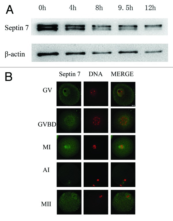
Figure 1. Subcellular localization and expression of Septin 7. (A) Samples were collected after oocytes had been cultured for 0, 2, 8, 9.5 and 12 h, corresponding to GV, GVBD, MI, AI and MII stages, respectively. The molecular mass of Septin 7 is 47 kDa and that of β-actin is 42 kDa. (B) Confocal microcopy showing immunostaining of Septin 7 (green) and DNA (red) in oocytes at GV, GVBD, pro-MI, MI, AI and MII stage. Bar = 10 μm.
