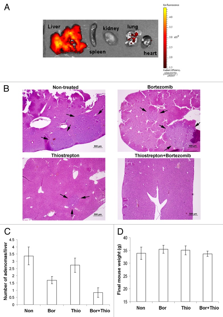
Figure 1. Analysis of hepatocellular tumor-bearing mouse livers after treatment with thiostrepton-micelles and/or bortezomib. Liver tumor models were prepared in FBV mice following the DEN/PB protocol, where neonatal pups were injected with DEN, followed by continuous exposure to PB by doping of drinking water. Thirty-two weeks post-DEN injection, treatment groups were administered with either 30 mg/kg thiostrepton-micelle, 0.5 mg/kg bortezomib or the combination of 30 mg/kg thiostrepton-micelle and 0.5 mg/kg bortezomib. Treatment continued for 5 weeks, after which animals were sacrificed and their livers prepared for histological examination. (A) Biodistributional studies of rhodamine-labeled micelle-thiostrepton (30 mg/kg) in liver tumor-bearing animals. Organ-associated fluorescence was examined 4 h post-administration using the Xenogen IVIS imaging equipment and demonstrated that the majority of micelle-thiostrepton accumulation was found in the liver of the animals. The spectral unmixing algorithm was used to differentiate between rhodamine and non-specific background autofluroescence. (B) Light microscopic evaluation of right liver lobe sections from control and treated animals. Black arrows point to the adenomas. (C) Post-treatment, the number of hepatocellular tumors in tissue sections was determined microscopically. Thiostrepton/bortezomib combination-treated animals exhibited significantly less number of adenomas compared with non-treated controls and single drug treated animals. (D) Thiostrepton-micelle, bortezomib and combination treatment did not cause significant weight loss in treated animals.

An official website of the United States government
Here's how you know
Official websites use .gov
A
.gov website belongs to an official
government organization in the United States.
Secure .gov websites use HTTPS
A lock (
) or https:// means you've safely
connected to the .gov website. Share sensitive
information only on official, secure websites.
