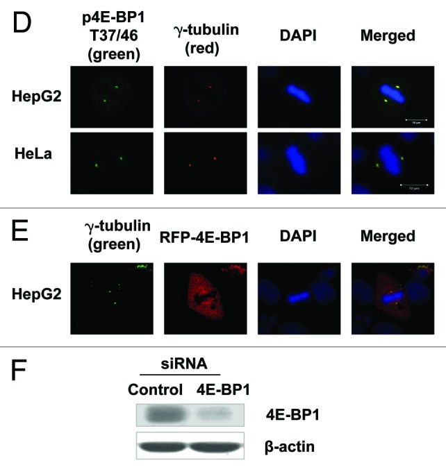
Figure 1D‒F. Phosphorylation of 4E-BP1 is linked with mitotic progression. (D) HepG2 (upper) and HeLa (lower) cells were immunostained with the antibody of phosphorylated 4E-BP1 /pT37/46 and γ-tubulin antibody and were examined by fluorescent microscopy. T37/46-phosphorylated 4E-BP1 overlapped with γ-tubulin at the centrosomes during mitosis. (E) HepG2 cells transfected with a vector of RFP-fused 4E-BP1 were immunostained with anti-γ-tubulin antibodies and subjected to fluorescent microscopy observation. RFP-fused 4E-BP1 protein was shown a diffuse distribution over the mitotic cell, but was more concentrated at the spindle organelle and co-localized with γ-tubulin at centrosomes. (F) The western blotting analysis of 4E-BP1 expression in HepG2 cells transfected with anti-4E-BP1 specific siRNA.
