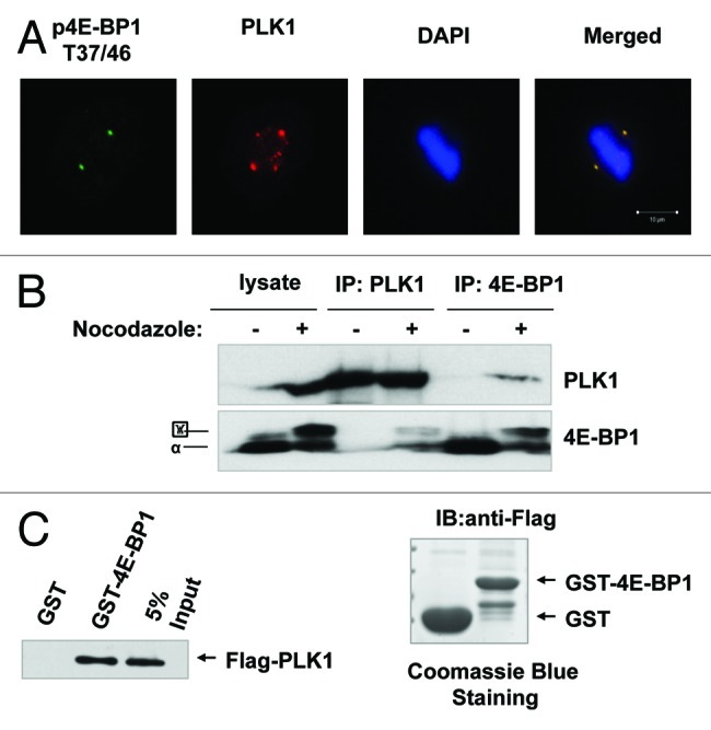
Figure 4A‒C. 4E-BP1 interacts with PLK1. (A) HepG2 cells were immunestained with antibodies against pT37/46 of 4E-BP1 or PLK1, and were examined by fluorescent microscopy. Phosphorylated T37/46 of 4E-BP1 co-localizes with PLK1 at centrosomes. (B) HepG2 cells were treated with nocodazole (200 ng/ml) for 16 h. Mitotic cells were collected by “shake-off.” Asynchronized and mitotic cells lysates were immunoprecipitated using anti-4E-BP1 or PLK1 antibodies. Western blot was performed using anti-PLK1 and 4E-BP1 antibodies. (C) GST and GST-fused 4E-BP1 recombinant bound to glutathione-Sepharose beads. 293T cells transfected with pCMV-2Btag-PLK1 lysates were incubated with the immobilized GST or GST-fusion protein. Bound fractions were analyzed by western blot using anti-Flag antibody.
