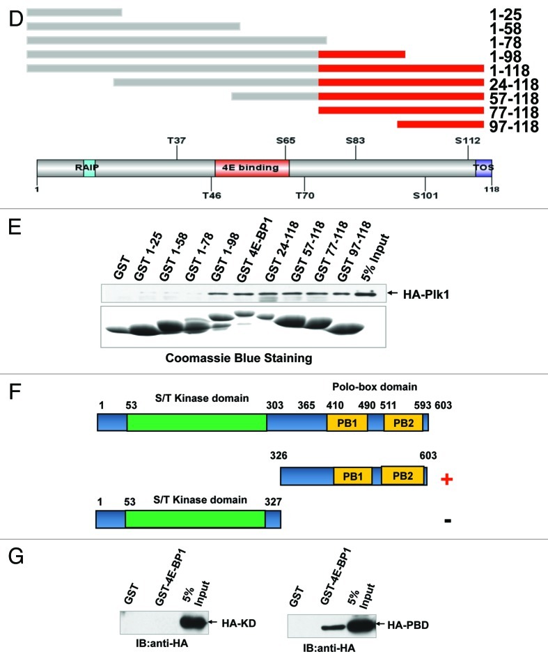
Figure 4D‒G. 4E-BP1 interacts with PLK1. (D) Schematic representation of 4E-BP1 protein deletion mutants. Red domain represents the interaction domain that mediates 4E-BP1 and PLK1 association identified in this study. (E) Recombinant GST and GST fused 4E-BP1 and its mutants bound to glutathione-Sepharose beads were incubated with 293T cells lysates which transfected with pCMV-HA-PLK1 vector. Bound fractions were analyzed by western blotting using anti-HA antibody. (F) Schematic representation of PLK1 protein deletion mutants. Design of these mutants was based on two main function domains of PLK1. Red minus represents the interaction domain. (G) Recombinant GST and GST-fused 4E-BP1 bound to glutathione-Sepharose beads. 293T cells transfected with pCMV-HA-PLK1 KD and PBD domain, respectively; after 48 h, cells were collected and lysates were incubated with the immobilized GST or GST-fusion protein. Bound fractions were analyzed by western blot using anti-HA antibody.

An official website of the United States government
Here's how you know
Official websites use .gov
A
.gov website belongs to an official
government organization in the United States.
Secure .gov websites use HTTPS
A lock (
) or https:// means you've safely
connected to the .gov website. Share sensitive
information only on official, secure websites.
