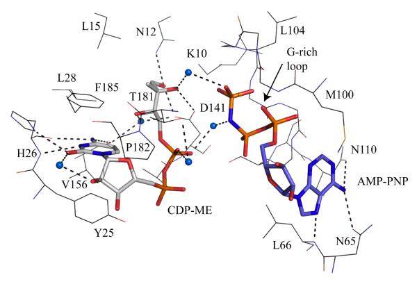
Figure 6.
The active site of E. coli IspE [47]. The IspE residues are shown as lines, the substrate and AMP-PNP as sticks using the same color scheme employed in Figure 6. Water molecules are blue spheres. Dashed lines represent potential hydrogen bonds.

