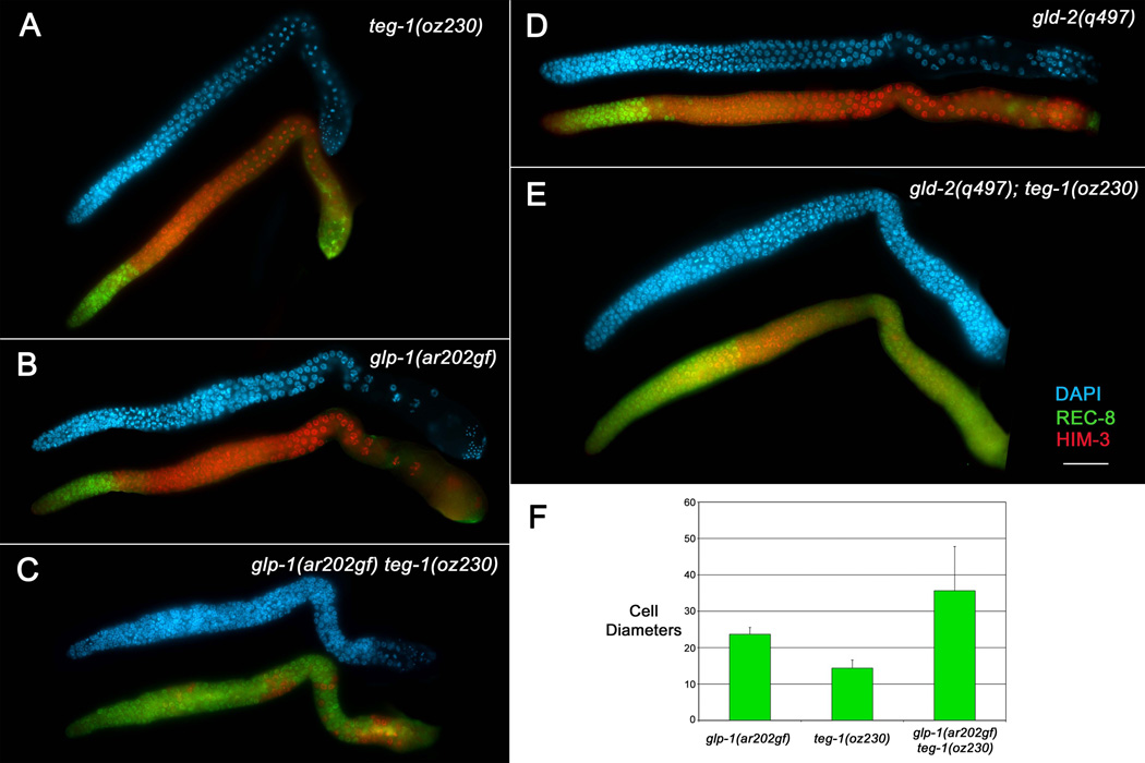
Figure 2.
teg-1(oz230) enhances the over-proliferative phenotype in different genetic backgrounds. Dissected gonad arms from adult hermaphrodite animals one day past the fourth larval stage. Gonad arms were stained with DAPI (blue) to show nuclear morphology, anti-REC-8 antibodies (green) to mark mitotic cells, and anti-HIM-3 antibodies (red) to mark meiotic cells. Both teg-1(oz230) (A) and glp-1(ar202gf) (B) animals, grown at 15°C, show proliferation restricted to the distal part of the gonad arm (although some anti-REC-8 cross-reactivity with sperm is observed in the proximal ends of the gonad arms); however, mitotic cells are found throughout the gonad arms of glp-1(ar202gf) teg-1(oz230) (C) animals (100% of gonad arms show this over-proliferation phenotype, n>20), although clusters of cells that have entered into meiosis are observed (red). (D–E) teg-1(oz230) also enhances the over-proliferative phenotype of gld-2(q497) single mutants (D), with gld-2(q497); teg-1(oz230) double mutants displaying extensive proliferation in both the distal and proximal regions of the gonad arms (E) (100% of gonad arms show this over-proliferation phenotype, n>20). All strains were also marked with unc-32(e189). Scale bar =20 microns. (F) The overall size of the of the distal mitotic zone is statistically larger in glp-1(ar202gf) teg-1(oz230) animals than in either single mutant, as measured by the number of cell diameters from the distal end of the gonad arm that are mitotic [anti-REC-8(+) (green) and distal to any meiotic nuclei -anti-HIM-3(+); (red)] (P < 9.7×10⁻⁵ t test. Error bars =1 SD. n= 20 gonad arms).

