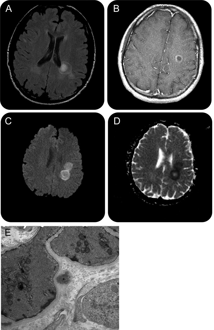
Figure. Brain MRI (A–D) and electron microscopy photomicrograph of skin biopsy (E).
(A–D) Concentric rings on brain MRI with restricted diffusion. (A) Axial brain MRI shows a lesion adjacent to the left lateral ventricle, which demonstrates a ring and central core hyperintensity on T2 fluid-attenuated inversion recovery image. (B) Axial T1 image of a gadolinium ring-enhancing lesion adjacent to the left lateral ventricle. (C) Axial diffusion-weighted image (DWI) shows high signal within 2 lesions. (D) Axial apparent diffusion coefficient map confirms the presence of restricted diffusion of lesions seen on DWI in (C). (E) Electron microscopy photomicrograph of skin biopsy: skin punch biopsy of the right deltoid was performed. A dermal arteriole with one definite granular osmiophilic material (GOM) deposit marked with an asterisk. The GOM deposit is in close apposition to the pinocytic vesicles of the vascular smooth muscle cell plasma membrane (magnification ×25,000).

