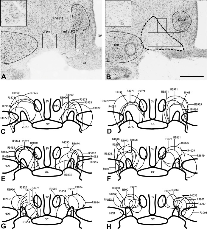
Figure 1.
Nissl stained brain sections from a rat that received saline into the VLPO (A) and a rat that received orexin-B-saporin into the VLPO (B). The outline of the lesion, marked by loss of large, darkly stained neurons and excess small glial nuclei, is shown in panel B (dashed line). This is better seen in enlarged views of the VLPO cluster from these sections in the corresponding insets. Counting boxes were placed over the VLPO cluster (VLPO), dorsal extended VLPO (dEVLPO), and medial extended VLPO (mEVLPO) as described in our previous study,24 and the number of remaining neurons were counted and then correlated with sleep and metabolic outcomes. Panels C-H show schematic representations of the location and extent of the lesions in each animal that received OX-SAP (n = 34). Note that a few animals had only unilateral lesions and are illustrated on only one side of the brain (R3898 in panel F and R3903 and R3860 in panel H). 3V, third ventricle; OC, optic chiasm; VLPO, ventrolateral preoptic area cluster; dEVLPO, dorsal extended VLPO; mEVLPO, medial extended VLPO; HDB, diagonal band of Broca horizontal; MPO, medial preoptic area. Scale bar = 500 μm.

