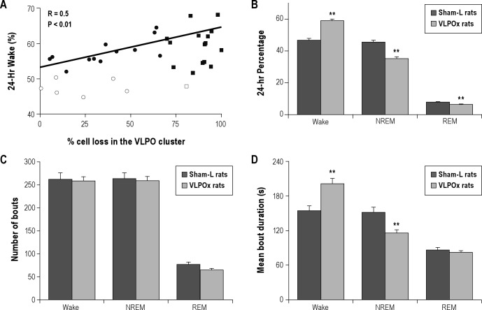
Figure 2.
Changes in sleep-wakefulness following lesions in the ventrolateral preoptic area (VLPO). Sleep-wake recordings were carried out on day 20 and on day 50 post-lesion from each animal and for 24 h on each occasion. As there were no significant differences in sleep-wake parameters between these 2 recordings, the mean values were calculated for each animal and were used for further analysis. (A) Daily percentage of wake in the entire cohort of animals where we attempted VLPO lesions was positively correlated with the percentage of bilateral cell loss in the VLPO. These animals were then classified in 2 ways—based on their cell loss in the VLPO cluster (> or < 70% cell loss) and based on sleep loss they experienced (> or < 10% sleep loss). Filled circles (•) = rats with < 70% cell loss in the VLPO but displayed > 10% sleep loss (n = 12). Open circles (∘) = rats with < 70% cell loss in the VLPO and had < 10% sleep loss (n = 6). Filled squares (■) = rats with > 70% cell loss and displayed > 10% sleep loss (n = 15). Open square (▫) = rat with > 70% cell loss in the VLPO but had < 10% sleep loss (n = 1). (B) Daily percentages of sleep-wake stages in sham-lesioned (sham-L; dark bars) animals (n = 7) and VLPO-lesioned (VLPOx; light bars) animals with > 70% cell loss (n = 16). VLPO lesions produced significant increase in wakefulness and a corresponding decrease in non-rapid eye movement sleep (NREM) and REM sleep. This sleep loss was brought about through an increase in average duration of wake bouts and a decrease in average duration of NREM sleep (D). The total number of wake, NREM or REM bouts during 24 h did not show any significant difference (C). Values are mean ± SEM **P < 0.01.

