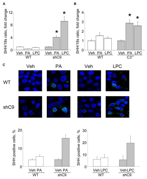
Figure 2. PA- and LPC-induces SHH expression in shC9 cells and primary mouse hepatocytes from caspase 3−/− animals.
(A) Wild type (WT) and shC9 cells were incubated with vehicle (Veh), PA at 800 μM for 16 hours, or LPC at 85 μM for 6 hours. SHH mRNA was quantified by real-time PCR, normalized to 18S rRNA, and expressed as fold change over vehicle. (B) Mouse primary hepatocytes (MPH) from WT and caspase 3 deficient animals (C3−/−) were incubated with Veh, PA at 400μM for 12 hours, or LPC at 42.5 μM for 6 hours. SHH mRNA was quantified by real-time PCR. (C) WT and shC9 cells were incubated with Veh, PA at 800 μM for 8 hours or LPC at 85 μM for 4 hours, respectively. Immunofluorescence was performed for SHH and visualized by confocal microscopy; the number of SHH-positive versus total cells is depicted. All data are expressed as mean ± SEM for three experiments; *p<0.01.

