Figure 4.
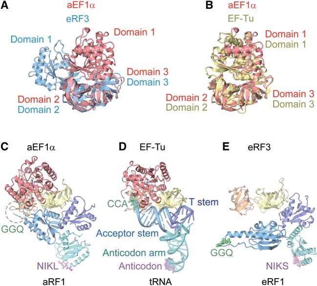
Comparison of the aRF1·aEF1α·GTP complex structure with related structures. (A) Superposition of domains 2 and 3 of aEF1α in the present complex (red) with those of the GDPNP-bound form of eRF3 (blue) (PDB ID: 1R5O). (B) Superposition of domains 2 and 3 of aEF1α in the present complex (red) with those of the GDPNP-bound form of EF–Tu (yellow) bound to tRNA (PDB ID: 1TTT). (C) The complex structure of aRF1·aEF1α·GTP (this work). Domains are color-coded as in Figure 1. (D) The complex structure of tRNA·EF–Tu·GDPNP (PDB ID: 1TTT). The domains of EF–Tu are color-coded as in aEF1α. The tRNA is colored turquoise (anticodon arm), light blue (acceptor stem) and purple (T stem). The bound GDPNP is depicted by a ball-and-stick model. (E) The complex structure of eRF1 and eRF3 lacking the GTP-binding domain (PDB ID: 3E1Y). Domains are color-coded as in aRF1·aEF1α.

