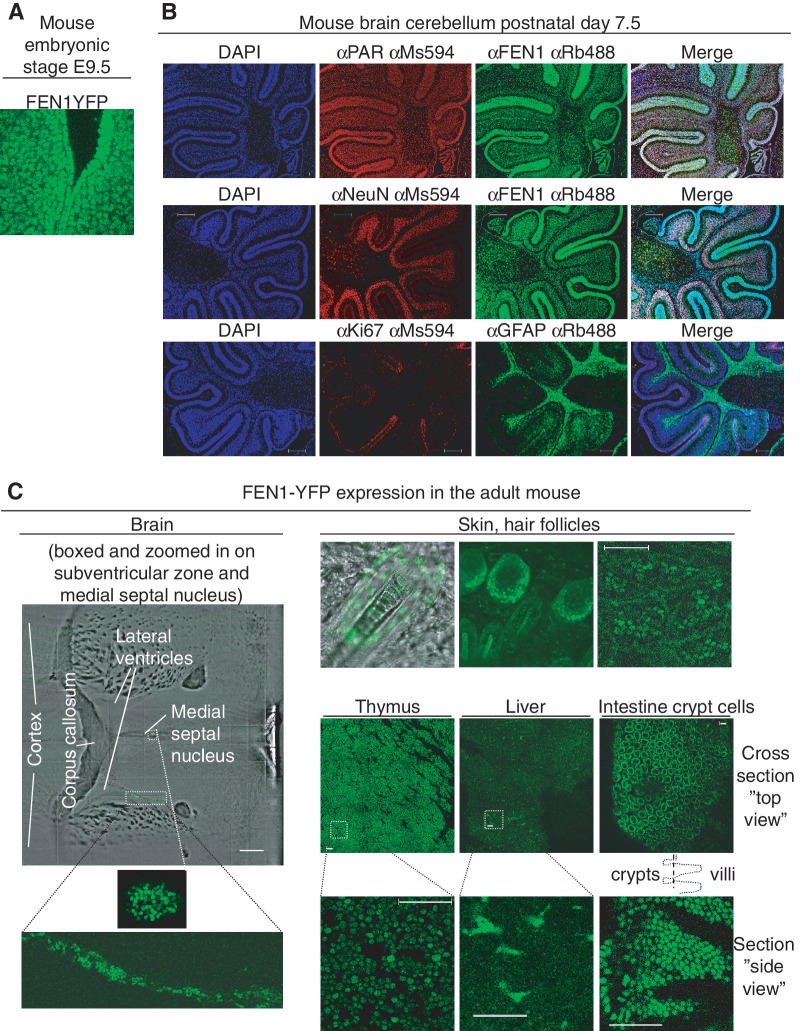
Figure 2.
FEN1-YFP expression in different mouse tissues. (A) In vivo imaging of FEN1-YFP expression. FEN1-YFP expression (green) is seen in the developing Fen1yNeo/yNeo mouse embryo (40×, embryonic stage E9.5, Confocal LSM 510 microscope). (B) Immunofluorescence staining of FEN1, poly (ADP-ribose) polymer (PAR, red in upper panel), brain cell markers (anti-NeuN stain neurons (red in middle panel) and anti-GFAP stain glia (green in lower panel)) and proliferation marker (anti-Ki-67, red in lower panel) in cerebellum from paraffin sections of Fen1y/y mouse brain 7.5 days after birth. Antibodies against the indicated polymer/proteins were used for co-staining of PAR and FEN1, NeuN and FEN1 and Ki-67 and GFAP. DAPI (blue) stained nuclear DNA. To the right, a merge of blue, red and green channel is shown. The scale bar is 200 μm. (C) Expression of FEN1-YFP in adult Fen1y/y mouse tissue. In brain from a 6 weeks old Fen1y/y mouse (left panel), FEN1-YFP cells could be detected in the subventricular zones and in the medial septal nucleus (Multiphoton LSM 710 microscope). Brain regions with FEN1-YFP expressing cells in the subventricular zone of one lateral ventricle and in the medial septal nucleus are enlarged (lower left panels). FEN1-YFP is expressed in skin (imaged with hair) from an adult Fen1yNeo/yNeo mouse (Confocal LSM 510 microscope), and in skin from an adult Fen1y/y mouse (Multiphoton LSM 710 microscope). In skin (three upper right panels), keratinocytes expressing FEN1-YFP could readily be detected in proliferative zones of the skin. FEN1-YFP expressing cells are readily found in thymus and intestine from a 6 weeks old Fen1y/y mouse (lower right panels). In liver from a 6 weeks old Fen1y/y mouse, FEN1-YFP expressing cells are found in a subpopulation of cells, which possibly could be Kuppfer cells, liver-specific macrophages (lower right panels). Kuppfer cells proliferate in the adult, and can have more than one nucleus. A Zeiss LSM 710 microscope with a 40× 1.3 oil objective was used for live imaging of FEN1-YFP in mouse tissue. The scale bar is 50 μm in images of live tissue, except for the 500 μm scale bar in brain.

