Figure 3.
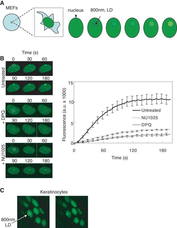
Mobility of FEN1-YFP in living cells. (A) Assay for local damage and fluorescence accumulation in MEFs. A circular area within the cell nucleus was irradiated with an 800 nm pulsed multiphoton laser to induce local DNA damage (dashed blue circle). Fluorescence in the locally damaged area was monitored over time. (B) FEN1-YFP accumulation after local damage. The PARP1-inhibitors DPQ and NU1025 was added to the indicated cells. FEN1-YFP showed a strong accumulation to the damaged site in untreated MEFs (n = 10), and a very weak accumulation to the region with DNA damage after addition of PARP1 inhibitors DPQ (n = 10) and NU1025 (n = 10) to the cells. The dashed white circle indicates the region in the cell nucleus where the DNA is damaged using a multiphoton laser at 800 nm. (C) Multiphoton laser at 800 nm was used to generate local DNA damage in keratinocytes of the Fen1y/y mouse skin, and FEN1-YFP was shown to accumulate in the regions with DNA damage. One representative image of skin cell nuclei before (left panel) and one image after (right panel) targeting with the multiphoton laser is shown (dashed white circle indicates damaged area of the targeted nucleus).

