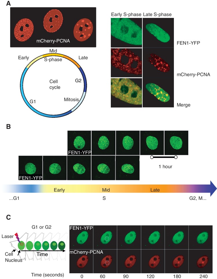
Figure 6.
FEN1-YFP dynamic behaviour throughout cell cycle and its colocalization with mCherry-PCNA. (A) Colocalization of FEN1-YFP and mCherry-PCNA in S-phase. Distinct PCNA replication structures are found in early, mid and late S-phase. In double colour MEFs expressing FEN1-YFP and mCherry-PCNA, PCNA appeared in very early S-phase and FEN1 foci could be detected soon thereafter (Multiphoton LSM 710 microscope). By late S-phase, perfect colocalization of FEN1 and PCNA foci was observed. (B) Live cell fluorescence imaging of FEN1 foci appearance and disappearance throughout S-phase. By mid S-phase distinct FEN1 foci were visualized, and the FEN1 foci were still present in late S-phase. By the end of late S-phase the FEN1 foci started to disappear (Multiphoton LSM 710 microscope). (C) Recruitment of FEN1 and PCNA to laser-induced DNA damage in G-phase cell. Non-S-phase MEFs expressing FEN1-YFP and mCherry-PCNA received irradiation with an 800 nm pulsed multiphoton laser (Multiphoton LSM 710 microscope). Fluorescence accumulation of FEN1-YFP and mCherry-PCNA to the DNA damage site was imaged over time. Early FEN1-YFP and mCherry-PCNA recruitment to the laser-induced DNA damage sites was found to be similar for both proteins.

