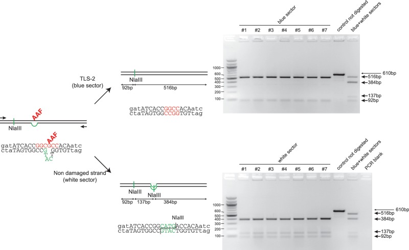
Figure 3.
Molecular characterization of blue and white sectors taken from lesion-containing sectored colonies. The sequence around the G-AAF adduct site shows the local heteroduplex that allows strand discrimination following replication. The non-damaged strand carries a 3 nt insertion that will generate an additional NlaIII restriction site during replication. Two PCR primers were designed to amplify a 610-bp fragment around the lesion site. The figure represents the analysis of individual sectors from seven sectored clones resulting from the integration of an AAF damaged vector (Figure 2B). Analysis of the white sectors (bottom gel) by NlaIII digestion shows the presence of an additional NlaIII restriction site corresponding to the replication of the non-damaged strand of the chromosome. Analysis of the blue sectors (top gel) lacks the additional NlaIII site; sequencing of these PCR fragments showed the expected sequence resulting from TLS events on the damaged strand (i.e. frameshift-2 in the NarI hotspot where the lesion is located (23).

