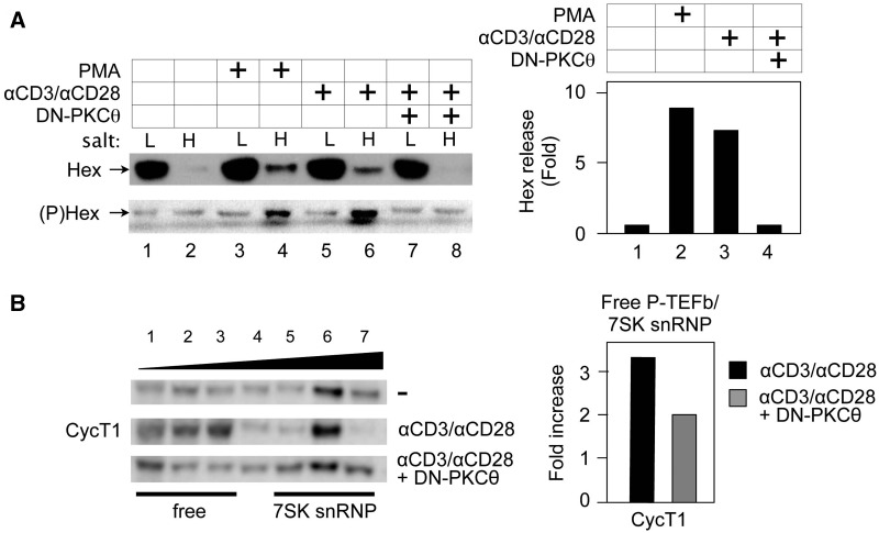
Figure 6.
Activation of PKC via phorbol ester or the TCR releases HEXIM1 in Jurkat T cells, which is blocked by the DN PKCθ protein. (A) PMA and anti-CD3/anti-CD28 antibodies release phosphorylated HEXIM1 protein from 7SK snRNP. Jurkat T cells expressed the empty plasmid vector (lanes 1–6) or DN-PKCθ (lanes 7 and 8). Forty-eight hours after the transfection, cells were untreated (lanes 1 and 2), or treated with PMA (20 nM, lanes 3 and 4), or anti-CD3/anti-CD28 antibodies (lanes 4 and 8) for 30min. High and low salt fractions were subjected to western blotting with anti-HEXIM1 (upper panel, Hex) and anti-phosphoylated HEXIM1 (lower panel, (P)Hex) antibodies. Hex release was determined as in Figure 5A. (B) The DN PKCθ protein inhibits the release of CycT1 from the 7SK snRNP. Left panel: Glycerol gradient sedimentation was performed as above (top panels, CycT1) and in the presence of DN-PKCθ (bottom panel). CycT1 of each fraction was detected with anti-CycT1 antibodies by western blotting. Whereas CycT1 in lanes 1–3 represents free P-TEFb, that in lanes 5–7 is in the 7SK snRNP. Right panel: Band intensities in fractions corresponding to free P-TEFb and 7SK snRNP were quantified and the ratio between these two groups was calculated and plotted as a fold-increase over the value obtained with unstimulated Jurkat cells.

