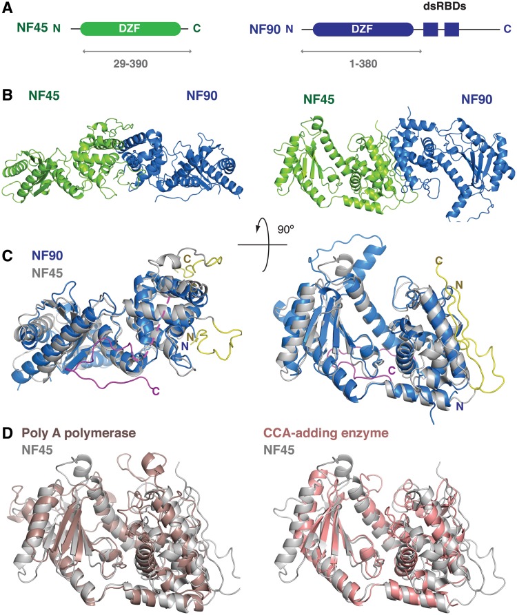
Figure 1.
Overview of the NF90/NF45 DZF dimerization domain structure. (A) Schematic representation of the domain organization of NF45 (green) and NF90 (blue). Double-headed arrows underneath indicate the sub-fragments of each protein that were used for crystallization. (B) Overview of the structure showing a ‘side’ view and a ‘top’ view rotated 90° around the horizontal axis. (C) Superposition of NF90 (blue) on NF45 (gray) using the same orientations of NF45 as shown in B. Extensions at the N- and C-termini of NF45 that are involved in dimerization interactions are shown in yellow. A C-terminal extension of NF90 is shown in magenta with a dotted line indicating a missing loop. (D) Superposition of NF45 (grey) on yeast Poly (A) polymerase (PDBid 2q66, brown) and CCA-adding enzyme from Archaeoglobus fulgidis (PDBid 1tfw, pink), shown from the ‘top’ view from (B).

