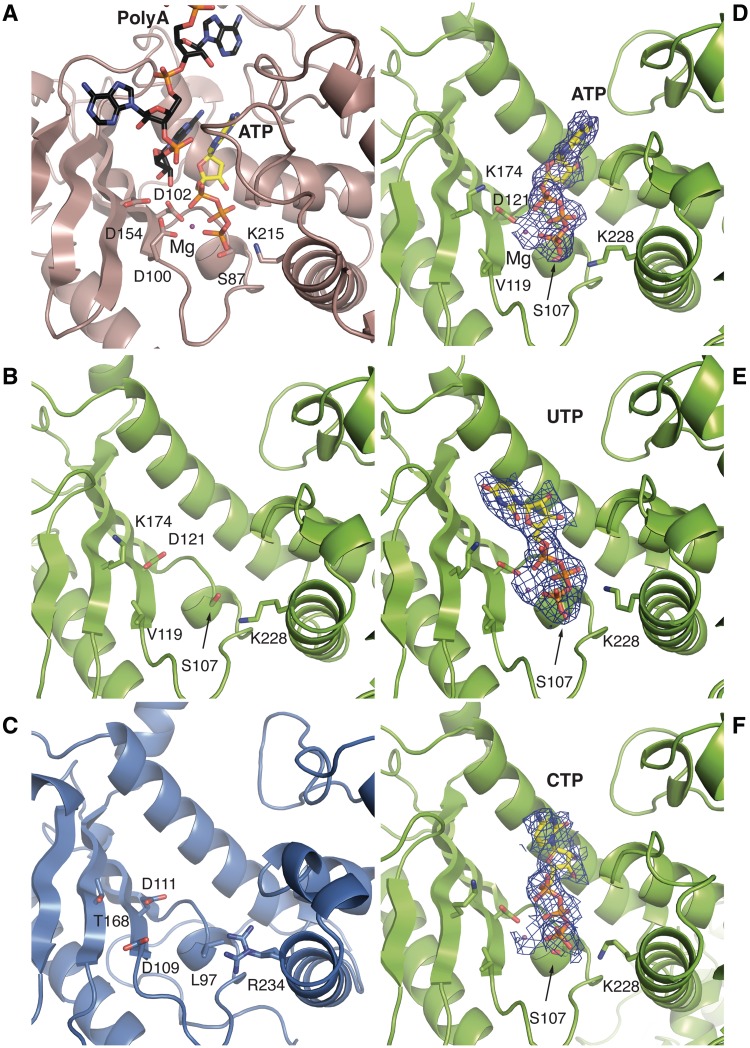
Figure 3.
A close-up view of the active site of poly (A) polymerase and equivalent regions in NF90 and NF45. (A) The catalytic cleft of yeast poly (A) polymerase (derived from PDBids 2q66 and 3c66) showing the catalytic residues and the bound ATP and a poly (A) substrate. (B) and (C) top view of NF45 and NF90 oriented as in Figure 1C, looking into the cleft between the two domains. The residues shown are those in equivalent positions to catalytic and nucleotide-binding residues found in related nucleotidyl transferases. (D) ATP bound to the cleft of NF45. A 2Fo–Fc map from the final refined structure is shown, contoured at 1σ. (E) and (F) A similar view as in (D) showing complexes with UTP and CTP, respectively.

