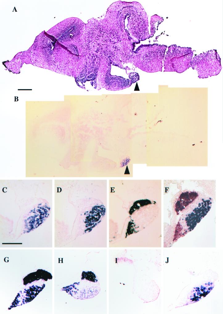
Figure 5.
In situ hybridization of sagittal sections of the pituitary (arrowhead) of stage-62 brains. (A) Hematoxylin and eosin stain. (B–J) In situ hybridization. (B) D2 in situ in a section adjacent to A. (C) Higher magnification of D2 in the pituitary in B. (D) TSHβ. (E) POMC. (F) Simultaneous in situ with POMC (orange) and TSHβ (purple). (G–J) Stage-58 tadpoles treated for 4 days with methimazole and iopanoic acid. (G and H) Simultaneous POMC and TSHβ in situ, but both probes were labeled with digoxigenin. (G) Stage-58 tadpoles treated for another 4 days with 10 nM T4. (H) Same as G, but treated for another 4 days with 10 nM T3. (I) D2 in situ of doubly inhibited stage-58 tadpole. (J) Same as I except treated for 4 days with 10 nM T4. (Bar for A and B = 200 μm.) (Bar for C–J = 100 μm.) In all figures, anterior is left and dorsal is up.

