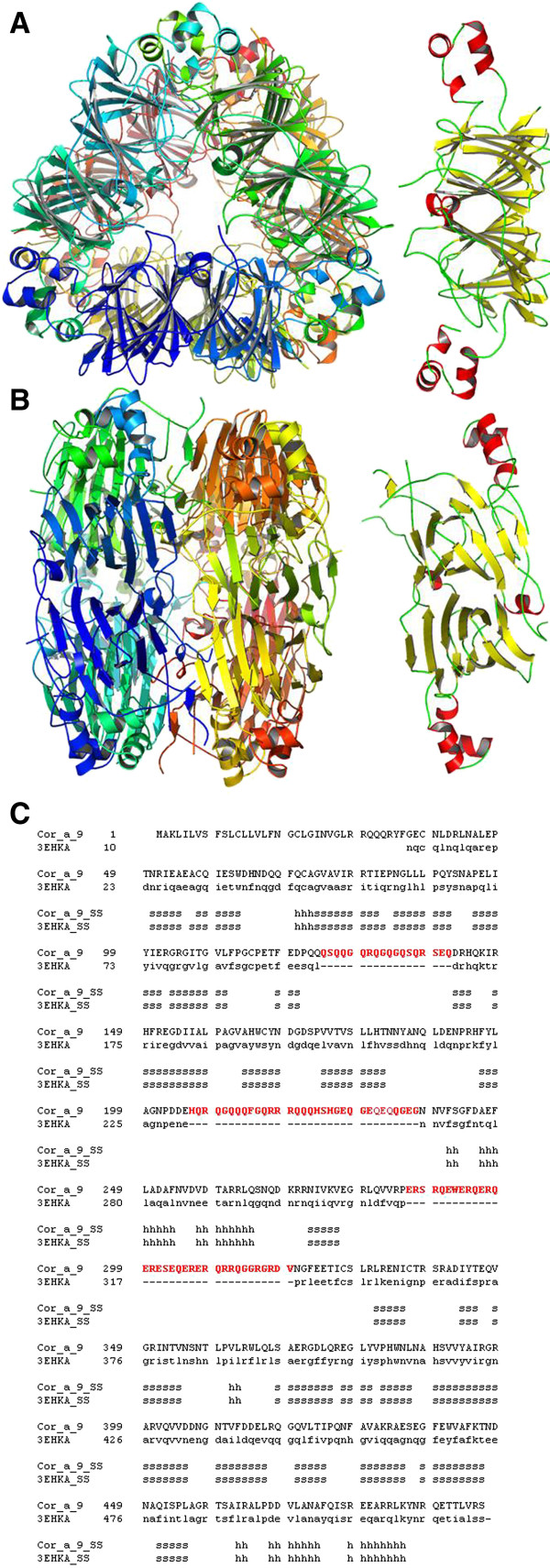
Figure 4.
3D structure model for Cor_a_9 homohexameric protein. (A). 3D model for Cor_a_9 hexamer (left) and single chain (right), top view. (B). Same as A, side view. (C). Structure alignment of Cor_a_9 protein sequence and the crystal structure of Pru du amandin from Prunus dulcis (PDB code: 3EHK; [37]). Secondary elements are indicated as in Figure1. Position of signal peptide is highlighted. Non-structured segments, absent in the template structure and therefore not modelled for Cor_a_9 sequence, are coloured in red.

