
Figure 1.
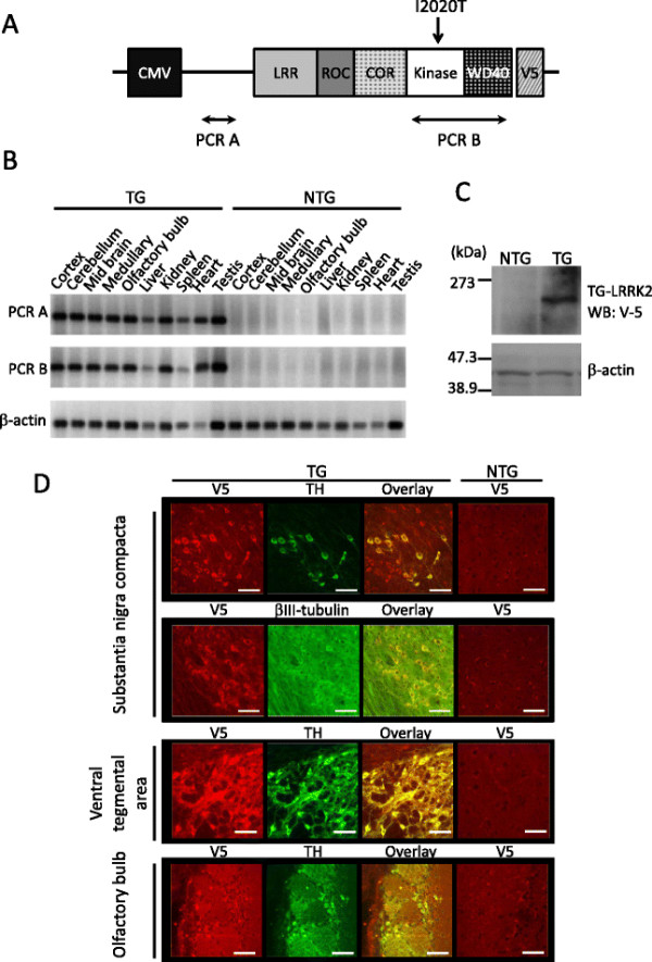
Expression of I2020T LRRK2 in the TG mouse. (A) Schematic representation of the LRRK2 cDNA transgene. (B) RT-PCR analysis of I2020T LRRK2 TG (line 41) and non-transgenic littermate mice. The amplified regions are shown in (A). (C) Western blotting analysis of whole brain. Lysates prepared from the whole brain of NTG and TG mice were subjected to Western analysis using anti V5-tag antibody. β-actin was used as a protein loading control. (D) Confocal immunostaining images of LRRK2 (V5), neurons (β-III tubulin), and dopamine neurons (TH) in the substantia nigra compacta, ventral tegmental area, and olfactory bulb of TG and NTG mice. Scale bar: 30 μm.