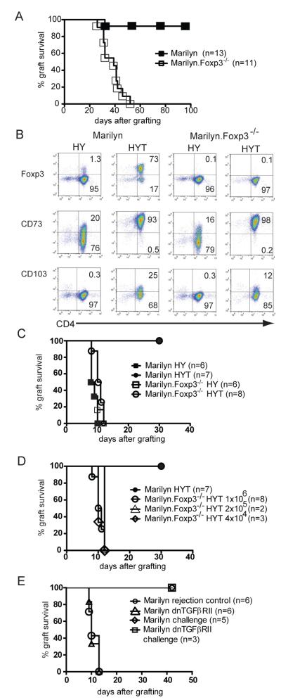
Figure 1. The relative requirements for Foxp3 and TGFß in induced tolerance to foreign antigen.
A. Female Marilyn mice (n=13) were transplanted with male B6.Rag−/− skin and treated with 1 mg of YTS177 (anti-CD4 blocking antibody) at days 0, 3 and 5 after grafting. Identical grafting and treatment was performed using female Marilyn.Foxp3−/− mice (n=11) and the grafts monitored for rejection (log rank p value <0.0001). Data are representative of two separate experiments.
B. Marilyn and Marilyn.Foxp3−/− splenocytes were cultured for 7 d with female B6 BMDC and HYAb peptide in the absence (HY) or presence (HYT) of TGFß. Histopaque-purified cells were co stained with CD4 and either Foxp3, CD73 or CD103 as shown. Numbers represent percentage of cells in the indicated quadrant. Data are representative of two separate experiments.
C. Marilyn or Marilyn.Foxp3−/− splenocytes were stimulated in vitro for 7 d with female B6 BMDCs and HYAb peptide in the absence (HY) or in the presence (HYT) of TGFß. One million Ficoll-purified live cells were injected into female B6.Rag−/− mice and one day later the mice were transplanted with a male B6.Rag−/− skin graft. Rejection was monitored daily. Data are representative of two separate experiments.
D. Using a similar experimental setting, the number of Marilyn.Foxp3−/− HYT splenocytes injected was titrated and is shown compared to the injection of 1×10⁶ Marilyn HYT cells (dark circles).
E. Female Marilyn mice made tolerant to male B6.Rag−/− skin by anti-CD4 antibody treatment (1 mg at days 0, 3 and 5 after grafting) were transplanted with a second male B6.Rag−/− skin graft 95-120 d after the first. At the time of the second graft the mice also received 1×10⁶ AutoMacs purified CD4+ T cells from female Marilyn or female Marilyn.dnTGFßRII mice and rejection was monitored daily. The rejection competency of the transferred Marilyn and Marilyn.dnTGFßRII T cells was validated by demonstrating the capacity of 1×10⁶ cells transferred into naïve female Rag−/− recipients to reject male Rag−/− skin grafts. The x-axis represents days after grafting primary grafts for the rejection controls, and days after second grafting for assessing the requirement for intact TGFß signalling in mediating dominant tolerance.

