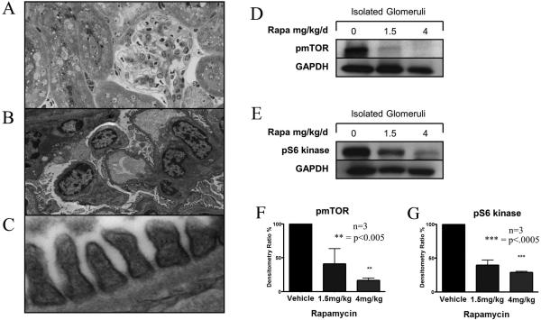
Figure 3. The treatment of mice with rapamycin inhibits glomerular expression of pmTOR and pS6 kinase.
C57BL/6 mice were treated with 1.5mg/kg/day or 4mg/kg/day of rapamycin i.p. or vehicle as a control. After 4 days, the kidneys were harvested and processed for light microscopy, electron microscopy and Western blot analysis. Panel A illustrates the histological appearance by light microscopy, and Panels B and C show the typical electron microscopy appearance of the renal cortex following treatment with rapamycin (4mg/kg). No histological changes are evident. Panels D-G show expression of pmTOR (Panels D, F), and pS6 kinase (Panels E, G) as evaluated by Western blot analysis in magnetic bead isolated glomeruli from control vehicle-treated and rapamycin-treated mice. Representative blots are illustrated in Panels D-E, and the mean densitometric analysis (+/- 1SD) from three different experiments are shown in Panels F and G. As illustrated, we found a dose-dependent reduction of glomerular expression of pmTOR and pS6 kinase in treated mice (vs. controls).

