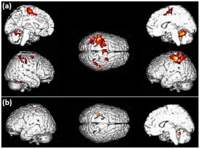
Figure 1. Patterns of brain activation during the hand movement task in PD patients (a – off L-dopa; b – on L-dopa).
Activation thresholds correspond to corrected (FWE) p-values<0.05. Without medication, brain activations were found in the right cerebellum, left motor/premotor regions, anterior cingulate cortex, superior and inferior parietal lobules, and putamen; following L-dopa intake, the brain activation profile was globally reduced, restricted to weak activations in the right posterior cingulate gyrus and the left inferior parietal lobule.

