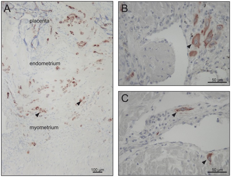
Figure 2. Invasion of extravillous trophoblast.
Immunohistochemistry using an antibody directed against cytokeratin 7 revealed the invasive depth and the numerical density of invasive cells in control cases (A, B) and early onset IUGR/PE (C). Original magnification 400× (B, C). (A) In order to allow publication of a survey picture illustrating the complete invasive pathway, immunoreactivity of cytokeratin 7-positive cells was enhanced by image analysis. Figure A represents the placental, endometrial and myometrial parts of the sample Original magnification 100×.

