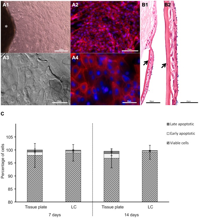
Figure 1. Cultivation and viability of LESCs.
Limbal graft (*) cultured on cell culture plate (A) or human LC (B) showing outgrowth of cells with epithelial morphology within 24 hrs of cultivation (image shown represents a 3 day cell outgrowth, A1 and A3 are bright field images, A2 and A4 are immunofluorescent images of actin cytoskeleton (red) and nucleus (blue)). Hematoxylin & Eosin staining of LESCs grown on LC (arrows) forming stratified epithelial layer at day 7 (B1 and B2). Cell viability and death of the cultured LESCs (viable cells (striped bar), early apoptotic or annexin V-FITC+ cells (light gray bar); late apoptotic or annexin V-FITC/propidium iodide+ cells (dark gray bar)) (C). Data shown are mean ± S.D (n = 3, Scale bars: 100 µm A1, 50 µm A2, 20 µm A3–4; 50 µm B1, 20 µm B2).

