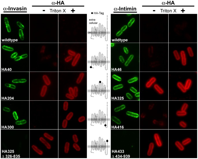
Figure 4. Immunofluorescence staining of invasin and intimin wild-type and HA-tagged variants expressed in E. coli omp2.
Bacteria were incubated with the primary antibody (anti-Inv, anti-EaeA or anti-HA). For detection of periplasmic protein the cells were permeabilized, as only epitopes that are exposed on the bacterial surface can be detected in non-permeabilized cells. Each sample was probed with antibodies directed against the C-terminal part of the passenger domain (C-terminal 195 aa of invasin or 280 aa of intimin, respectively) and antibodies directed against the HA tag. Miniaturized invasin topology models with the insertion sites of the HA tag (black dots) are shown in the middle of the figure. As the predicted position of the labels also applies for intimin and for sake of clarity, we did only include the invasin miniature models in this figure.

