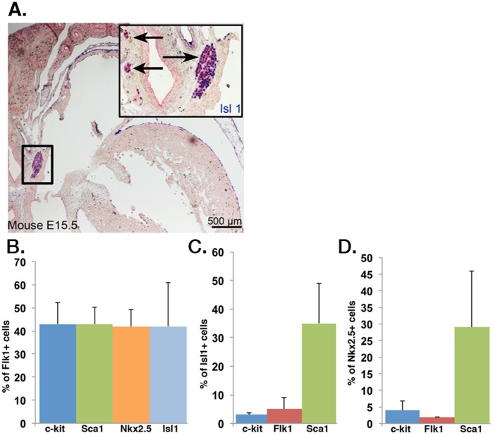
Figure 1. Flk1 is not a specific marker for endogenous and mouse ESC-derived Isl1+ CPCs.
(A) Immunohistochemical staining of E15.5 mouse heart identifies Isl1-expressing CPCs (blue) located in niches in outflow tracts. (B) FACS analysis of mouse ESC-derived Flk1+ cells reveals a heterogeneous Flk1+ population with low enrichment for Isl1 (light blue bar) and Nkx2.5 (orange bar) cells (n = 3). (C & D) FACS analysis of differentiated mouse ESCs reveals that Flk1 represents <10% of Isl1+ cells (C; n = 3) and <5% of Nkx2.5+ cells (D; n = 3).

