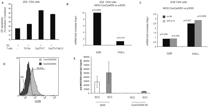
Figure 6. Induction of cytolytic factors in CD4 T cells stimulated with CxxC modified peptides.
(A) CD4+CD62L+ cells from 2D2 transgenic mice stimulated for three cycles under Th17 polarizing conditions with wt or modified peptide (as in Figure 5) were added to LPS activated dendritic GFP-transduced JAWS II cells (ratio T/DC: 2/1) loaded with peptide MOG 35–55. After 20 h, apoptosis of JAWS cells was measured by Annexin V staining of GFP positive cells. Error bars respresent 1 SD. (***p = 0.0018; NS p = 0.02). Two tailed p values are from unpaired t test. (B) quantitative PCR for FAS-L and GZB on polyclonal 2D2 CD4 cells differentiated under TH17 conditions with wild type or CxxC peptide (same cells as in Figure 5A). Fold change for CxxC-GG40–55 cells relative to MOG35–55 generated cells was determined after stimulation with APC loaded wild type MOG35–55. Results were normalized to 18S rRNA. Two tailed p values are derived from unpaired t tests. (C) Quantitative PCR on EAE CD4 cells after 3 cycles of in vitro amplification (in the presence of IL-23 and TGF–β) with MOG35–55 or CxxCGG40–55. Fold change for CxxCGG40–55 cells relative to MOG35–55 generated cells was determined after stimulation with T cell depleted splenocytes loaded wild type 35–55. Grey histograms are for CxxCGG40–55 stimulated in the presence of an anti-IL-12 antibody. Results were normalized to 18S rRNA. Results are representative of two independent experiments. Two tailed p values are derived from unpaired t tests. (D) CD4+CD62L+ cells from 2D2 transgenic mice were expanded for three cycles with CxxCGG40–55 or loss of function SxxSGG40–55. Ten days after the third stimulation, cells were stimulated with MOG 35–55 for 18 h and stained for intracellular GZB. Grey histogram is for cells expanded with CxxCGG40–55 and black histogram for SxxSGG40–55 expanded cells. Open histogram is for isotype control staining (iso). Data representative of two experiments. (E) C57BL/6 mice were immunized by 3 footpad injections of 10 µM MOG3555 or CxxCGG40–55 peptides in Incomplete Freund’s adjuvant at 11 days intervals. Ten days after the second (SC2) and third (SC3) injections, mice (n = 5 in each group) were bled and the presence of anti-MOG35–55 IgG antibodies was detected in a quantitative Elisa assay.

