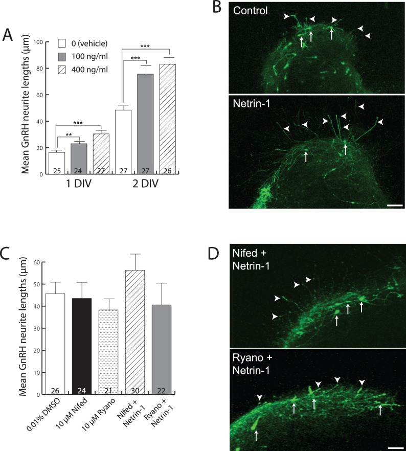
Figure 2. Netrin-1 stimulates the growth of GnRH neurites in a calcium-dependent manner.
(A) Bar graph showing mean GnRH neurite lengths at 1 and 2 DIV across the three different treatments (indicated in inset legend). Numbers at the base of each bar indicate the number of explants used, and are from fetal brains derived from 5 independent pregnancies (average of 8 fetuses/pregnancy). Error bars indicate SEM. **, P<0.01 and ***, P<0.001 with one-way ANOVA and Student-Newman-Keuls multiple comparison post-hoc test. (B) Immunofluorescence images for detection of GFP peptide as a surrogate marker for GnRH neurons (arrows) of explants grown for 2 DIV. Note the clear difference in the length of GnRH neurites (arrowheads) outside the explant borders in Netrin-1-treated (lower panel) by comparison with untreated (upper panel). Scale bar, 40 µm. (C) Bar graph showing mean GnRH neurite lengths (y-axis) at 2 DIV under the three treatment conditions indicated on the x-axis. Numbers at the base of each bar indicate the number of explants used, and are from fetal brains derived from 4 independent pregnancies. Abbreviations: Nifed = nifedipine, Ryano = ryanodine. Error bars indicate SEM. (D) Immunofluorescence images showing GFP-expressing GnRH neurons grown in explants in the presence of Netrin-1 plus either the L-type VGCC blocker Nifedipine (Nifed + Netrin-1, upper panel) or ryanodine (Ryano + Netrin-1, lower panel). Arrows indicate GnRH neuron cell bodies, and arrowheads indicate GnRH nerve fibers extending beyond the borders of the explants. Scale bar, 100 µm.

