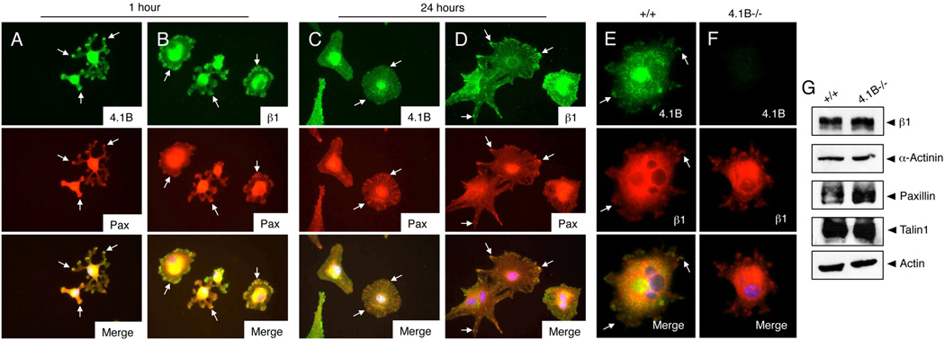
Figure 2. Protein 4.1B displays dynamic sub-cellular localization in spreading astrocytes.
(A–D); Wild type astrocytes were plated on fibronectin-coated dishes for 60 minutes (A, B) or 24 hours (C, D). Spreading cells were then labeled with anti-4.1B (A, C) or anti-β1 integrin (B, D) rabbit polyclonal antibodies in combination with anti-paxillin mAbs. (E, F); Wild type (E) and 4.1B−/− (F) astrocytes were plated on fibronectin for one hour and then immunolabeled with anti-4.1B polyclonal antibodies and anti-β1 integrin mAbs. Protein 4.1B and β1 integrin co-localize at one hour post-adhesion on fibronectin (arrows in E). Images are shown at 400×. (G); No differences in expression of common focal adhesion proteins in wild type and 4.1B−/− astrocytes.

