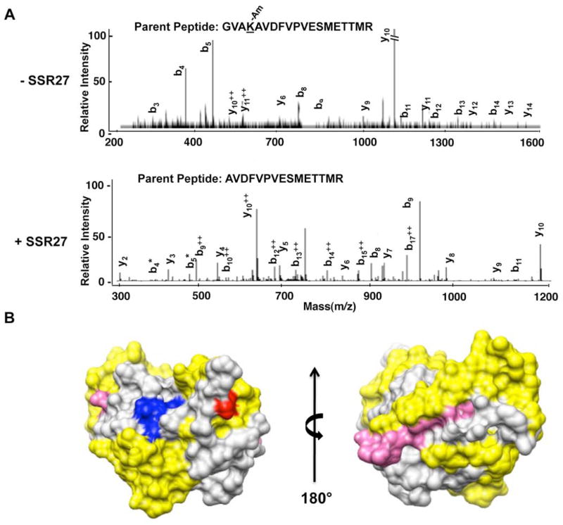
Fig. 2.
Amidination of lysine residues in the NS3P domain in the absence and presence of RNA.
A) MS/MS analysis of peptides from NS3P that were differentially amidinated in the presence and absence of RNA. The top panel shows the b and y ions from the MS/MS spectra for the peptide that was not cleaved by trypsin due to amidination at Lysine 165. The bottom panel shows the MS/MS spectra for the parent peptide in which amidination did not take place due to the presence of ssR27, and the peptide was cleaved at the cognate lysine. B) The model for NS3P and the location of the RCAP peptides relative to the active site of NS3P. The active site residues are in blue. The locations of peptides identified to crosslink to RNA in Fig. 1 are in yellow, and the location of the amidinated lysine (K165) is in red.

