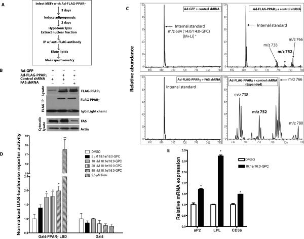
Figure 4. Isolation of FAS-dependent Diacyl and 1-O-alkyl Ether Phosphatidylcholine Species Associated with PPARγ.
(A) Strategy for detection of PPARγ-associated lipids.
(B) Detection of FLAG-PPARγ protein immunoprecipitated from adipocytes treated with control or FAS shRNA.
(C) Mass spectrometric analyses of [M+Li]+ ions of glycerophosphocholine (GPC) lipids bound to FLAG-PPARγ or control protein (GFP) immunoprecipitated from control and FAS knockdown adipocytes. Ions of m/z 752 and 780 represent 1-O-alkyl GPC species.
(D) CV-1 cells were transfected with plasmids encoding UAS-luciferase, Renilla luciferase and Gal4-PPARγ LBD (a C-terminal fragment of PPARγ encompassing the ligand binding domain fused to the Gal4 DNA binding domain) or Gal4 alone. The cells were treated with 18:1e/16:0-GPC (corresponding to m/z 752 in panel C), rosiglitazone or DMSO. After 48 hrs, UAS-luciferase reporter activity was measured and normalized to Renilla luciferase reporter activity. **P=0.0001. *P=0.018 (10 μM), 0.024 (20 μM), 0.019 (80 μM).
(E) 3T3-L1 cells were induced to differentiate in DMEM+10% FBS with supplemental dexamethasone, insulin and IBMX in the presence of 20 μM 18:1e/16:0-GPC or DMSO. After 3 days, the cells were re-treated with the GPC in media containing supplemental insulin alone. The next day, the cells were harvested for RNA extraction and real-time PCR analysis. The data are representative of 3 separate experiments. *P=0.0147 (aP2), 0.0006 (LPL), 0.0102 (CD36).
Error bars in panels D and E represent SEM.

