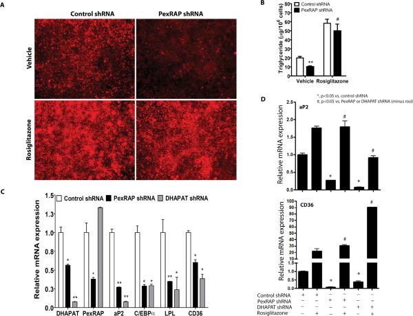
Figure 6. PexRAP is Required for Adipogenesis and PPARγ Activation.
(A) Nile red staining of 3T3-L1 adipocytes treated with control or PexRAP shRNA in the presence or absence of rosiglitazone.
(B) Triglyceride content for the cells of panel A. **P=0.0066 vs. control, #P=0.0071 vs. PexRAP shRNA vehicle. N=3/condition.
(C) RT-PCR analysis of gene expression following PexRAP or DHAPAT knockdown. P vs. control: DHAPAT, *0.0278, **0.007; PexRAP, *0.040; aP2, **0.0060 for PexRAP shRNA and 0.0058 for DHAPAT shRNA; C/EBPα, *0.0160 for PexRAP shRNA and 0.0165 for DHAPAT shRNA; LPL, **0.0014, *0.0450; CD36, *0.0113 for PexRAP shRNA and 0.0132 for DHAPAT shRNA. N=3–5/condition.
(D) Rosiglitazone treatment rescues the effect of PexRAP or DHAPAT knockdown on PPARγ target gene expression. 3T3-L1 cells infected with lentivirus expressing control, PexRAP, or DHAPAT shRNA were induced to differentiate into adipocytes and then treated with 2.5 μM rosiglitazone. Expression of PPARγ target genes was analyzed by quantitative RT-PCR. For aP2, exact P values from left to right= 0.0038, 0.0119, 0.0024, 0.0032. For CD36, P values= 0.0022, 0.0015, 0.0110, <0.0001.
Error bars in panels B–D represent SEM.

