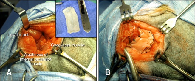
Fig. 2.

Intraoperative photographs of perineal herniorrhaphy using canine small intestinal submucosa (SIS) in Case 1. (A) The four-layered canine SIS sheet was prepared. After placing the deviated rectum in its original location, the levator ani, coccygeus, and internal obturator muscles were identified. (B) The canine SIS sheet was placed between the perineal muscles.
