Figure 4.

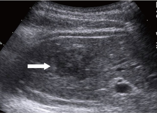
Sonographic finding. The lesion (arrow) is a well-circumscribed, hypoechoic mass compared with surrounding liver parenchyma.

Sonographic finding. The lesion (arrow) is a well-circumscribed, hypoechoic mass compared with surrounding liver parenchyma.