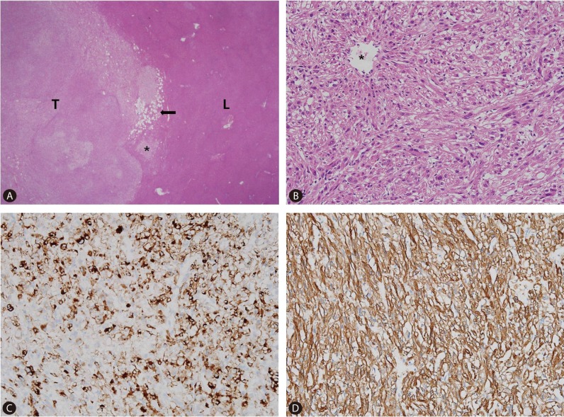
Figure 6.
Histologic and immunohistochemical findings. (A) Tumor (T) and liver parenchyma (L) have relative good delineated interface with focal infiltrative margin (asterisk). Fat vacuoles are aggregated in the periphery of the tumor (arrow), but are very rarely identified in the center (original magnification ×40). (B) The spindle shaped tumor cells are arranged in short intersecting fascicles. The cells have abundant eosinophilic cytoplasm and ill-defined cell membrane. (C) Nuclei are round to oval and have small nucleolus. The tumor cells grow radiating from the wall of vessel (asterisk). The spindle cells are positive for human melanocyte B-45 (HMB-45). (D) Smooth muscle actin. HMB-45 is strongly positive in this tumor. Smooth muscle actin demonstrates diffuse strong positive staining in the tumor cells.

