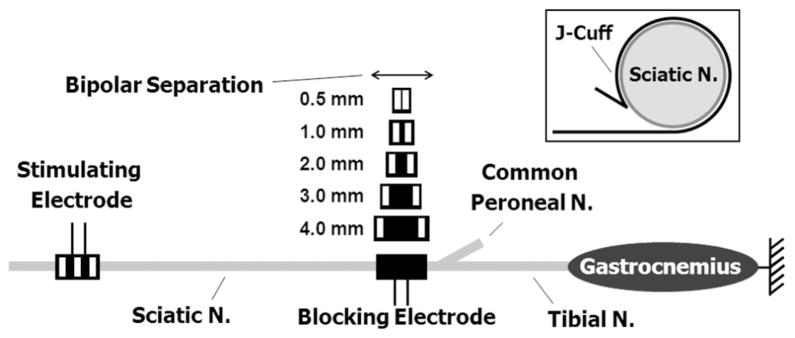
Fig. 2.
Diagram of the experimental setup showing nerve cuff electrode placement. A tripolar proximal stimulating electrode was always used. HFAC blocking nerve cuff electrodes of various geometries were placed at the branch point for the common peroneal nerve for each of the block randomized trials. The inset shows a cross sectional depiction of a J-cuff electrode on the sciatic nerve.

