Abstract
Aim:
Supernumerary teeth are considered as one of the most significant dental anomalies during the primary and early mixed dentition stage. The main objective of the study was to determine the prevalence rate of supernumerary teeth in the patients who reported to the Department of Oral Medicine and Radiology and to study the associated clinical complications.
Materials and Methods:
A longitudinal observational study was conducted of 2216 patients for a period of 4 months with the documentation of demographic data, the presence of supernumerary teeth, their location, and associated complications such as mechanical trauma, dental caries, and associated pathology.
Results:
The study recorded 27 supernumerary teeth from the examined 2216 patients. This yields a prevalence of 1.2%, with greater frequency in males which was 1.49% and in females the frequency was 0.85%. The greatest proportion of supernumerary teeth was found in the maxillary anterior region (77.8%). Out of this, 85.7% were classified as mesiodens based on their location. The displacement of adjacent teeth was the most common finding, followed by dental caries.
Conclusion:
The prevalence of supernumerary teeth in this study was 1.2% which is in agreement with that reported in similar studies and the maxillary mesiodens was the most common location. Displacement of adjacent teeth was the most common finding.
KEY WORDS: Displacement, maxillary mesiodense, supernumerary teeth
A supernumerary or hyperdontia tooth describes an excess in teeth number which can occur in both the primary and the permanent dentition.[1,2] Its prevalence is higher in males than females (2:1).[3] Depending on the literature source, the reported frequency in permanent dentition varies from 0.1 to 3.6% in the general population[4,5] and a few cases of inherited forms have been reported.[6,7] There is no commonly accepted etiology for supernumerary teeth, but hyperactivity of the dental lamina is the most widely accepted theory.[1,8] In some cases, there appears to be a hereditary tendency for the development of supernumerary teeth.[1] Generally, supernumerary teeth may erupt in any part of the dental arch, but they most commonly occur in the maxillary midline region, i.e. mesiodens, which wis approximately 80% of all supernumerary teeth.[9,10] The second common supernumerary tooth is the maxillary fourth molar (distomolar) and is situated distal to the third molar.[1] A mandibular distomolar is also seen occasionally, but this is much less common than the maxillary distomolar.[1] Other supernumerary teeth seen with some frequency are maxillary paramolars situated buccally, lingually, or interproximally in molar areas, mandibular premolar, and maxillary lateral incisors.[1] Supernumeraries in the region of mandibular central incisors and maxillary premolars are found occasionally.[1]
In comparison to permanent dentition, the frequency of occurrence of supernumerary teeth in primary dentition is five times lower.[11–13] Clinically, the supernumerary teeth are usually coincidently diagnosed during either intraoral or radiograph examination. Supernumerary teeth may cause different local disorders, including crowding, disturbed eruption, or retention of teeth, delayed or abnormal root formation in permanent teeth, and cyst.[14]
The extraction of these teeth is a general rule for avoiding complications,[15] but some authors having different views. For example, Koch et al.[16] do not recommend extractions of impacted teeth in children under 10 years of age, since in this particular age group, such procedures often require general anesthesia. The purpose of this study was to determine the epidemiological characteristics of supernumerary teeth, with an analysis of the associated clinical eruptive complications.
Materials and Methods
A longitudinal, observational study was conducted in 2216 patients who reported to the Department of Oral medicine and Radiology (KSRIDSR, Tamilnadu, India), to identify the frequency of supernumerary teeth occurrence. For each patient with supernumerary teeth, we recorded the demographic variable (age and sex) following clinical examination. In required situations, we took intraoral periapical radiograph (IOPA) to confirm the condition and we documented the location of the tooth. The pathology associated with the supernumerary teeth was also recorded, such as displacement, delayed eruption, dental caries, and associated lesions (presence of follicular cyst). All obtained data were statistically analyzed with SPSS-16.0 version (SPSS, Inc, Chicago, IL, USA) software program for windows by using descriptive statistics, cross tabulations, and chi-square test.
Results
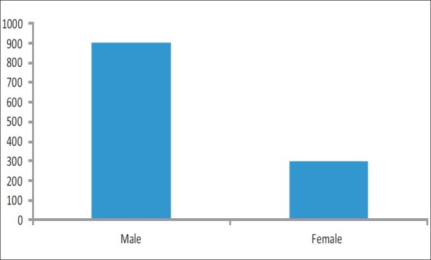
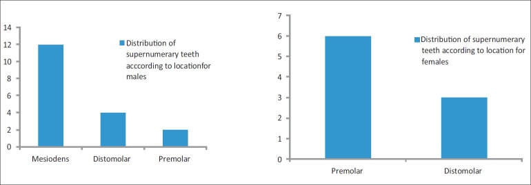
Twenty-seven supernumerary teeth were detected from 2216 patients examined [Table 1]; this yields a total prevalence of 1.2%, 1.49% in males, and 0.85% in females [Table 2, Figure 1]. In the total 27 supernumerary teeth observed, 18 (66.7%) were located in the maxilla and 9 (33.3%) in the mandible. In maxilla, the most common supernumerary teeth was mesiodens (44.44%, n = 12) followed by distomolar (14.81, n = 4) and in premolar region was (7.4%, n = 2) [Tables 3 and 4, Figure 2]. In mandible, the most common location was premolar region (22.22%, n = 6) followed by distomolar (11.11, n = 3) [Table 5, Figure 2]. The prevalence of supernumerary tooth was nil in the maxillary canine and mandibular incisors and canine region. The overall prevalence rate of supernumerary teeth was higher in the maxillary anterior region (51.85, n = 14) in comparison to the other areas, followed by mandibular premolar region (22.22%, n = 6). In comparison to the other areas (22.2%) of maxilla, supernumerary teeth were found commonly in the anterior region (77.8%). In mandible, the most frequent location was premolar region (66.67%) than the other locations (33.33%). The mean age of the patients with supernumerary teeth was 24.6 years. The teeth most commonly manifested in the 3rd decade of life (46.3%), followed by the 2nd decade (29.2%).
Table 1.
Number of patients examined

Table 2.
Supernumerary teeth found

Figure 1.

Frequency of supernumerary teeth occurrence among sexes
Table 3.
Distribution of supernumerary teeth

Table 4.
Distribution in maxilla

Figure 2.
Distribution of supernumerary teeth according to location
Table 5.
Distribution in mandible

Discussion
Based on the study findings, the prevalence of supernumerary teeth in 2216 patient was 1.2%, which coincides with the other studies, with a range of 0.1–3.8%.[17,18] The mean age of the patients in our study was 24.6 years, i.e., 3rd decade of life. It coincides with the findings of other authors who report this decade to be the most common period for presence of supernumerary tooth. In terms of location, most commonly, the supernumerary teeth occur in the maxillary anterior region.[19] The present study is in agreement with this report, as 77.8% was found in the anterior maxilla.
Regarding gender distribution, most of the supernumeraries in this study showed that the prevalence was much higher among males (1.49%) than females (0.85%), i.e., the ratio coincides with that in other studies.[18,20–23] Considering the mechanical and pathological changes induced by supernumerary teeth, displacement of adjacent teeth is higher (40.7%), followed by dental caries, which coincides with the findings of Leco-Berrocal MI et al.[24]
Prevalence rate of supernumerary teeth among the non-syndromic South Indian population – An analysis
Total number of patients examined: 2216
Age group: 7–32 years
Mean age: 24.6 years
Conclusion
The prevalence of supernumerary teeth in the patients who reported to the dental OP was 1.2% and is comparable to that reported in previous studies. The most common area for supernumeraries is maxillary anterior region. The most common complication found in association with supernumeraries is displacement of adjacent teeth.
Footnotes
Source of Support: Nil
Conflict of Interest: None declared.
References
- 1.Shafer WG, Hine MK, Levy BM, editors. Textbook of oral pathology. 6th ed. Noida: Elsevier publication; 2009. pp. 46–7. [Google Scholar]
- 2.Dominguez A, Mendoza A, Fernandez H. Estudio rettrospetivo de dientes supernumeraries en 2045 pacientes. Av Odontoestomatol. 1995;11:575–82. [Google Scholar]
- 3.Sharma A. Familial occurrence of mesiodens-a case report. J Indian Soc Pedod Prev Dent. 2003;21:84–5. [PubMed] [Google Scholar]
- 4.Acikgoz A, Acikgoz G, Tunga U, Otan F. Characteristic and prevalence of non-syndrome multiple supernumerary teeth: A retrospective study. Dentomaxillofac Radiol. 2006;35:185–90. doi: 10.1259/dmfr/21956432. [DOI] [PubMed] [Google Scholar]
- 5.Zhu JF, Marcushamer M, King DL, Henry RJ. Supernumerary and congenitally absent teeth. A literature review. J Clin Pediatr Dent. 1996;20:87–95. [PubMed] [Google Scholar]
- 6.Yusof WZ, Awang MN. Multiple impacted supernumerary teeth. Oral Surg Oral Med Oral Pathol. 1990;70:126. doi: 10.1016/0030-4220(90)90190-4. [DOI] [PubMed] [Google Scholar]
- 7.Burzynski NJ, Escobar R. Classification and genetics of numeric anomalies of dentition. Birth Defects Orig Artic Ser. 1983;19:95–106. [PubMed] [Google Scholar]
- 8.Primosch RE. Anterior supernumerary teeth-Assessment and Surgical Intervention in children. Pediatr Dent. 1981;3:204–15. [PubMed] [Google Scholar]
- 9.Alaejos C, Contreras MA, Buenechea R, Berini L, Gay C. Mesiodens: A retrospective de una serie de 44 pacientes. Med Oral. 2000;5:81–8. [PubMed] [Google Scholar]
- 10.Danalli DN, Buzzato JF, Braum TW, Murphy SM. Long-term interdisciplinary management of multiple mesiodens and delayed eruption: Report of a case. J Dent Child. 1998;55:376–80. [PubMed] [Google Scholar]
- 11.Rajab L, Hamdan M. Supernnumerary teeth: Review of the literature and a survey of 152 cases. Int Jof Pediatr Dent. 2002;12:244–54. doi: 10.1046/j.1365-263x.2002.00366.x. [DOI] [PubMed] [Google Scholar]
- 12.Sedano H, Gorlin R. Familial occurrence of mesiodens. Oral Surg Oral Med Oral Pathol. 1969;27:360–1. doi: 10.1016/0030-4220(69)90366-1. [DOI] [PubMed] [Google Scholar]
- 13.Sykaras S. Mesiodens in primary and permanent dentitions. Report of a case. Oral Surg Oral Med Oral Pathol. 1975;39:870–4. doi: 10.1016/0030-4220(75)90107-3. [DOI] [PubMed] [Google Scholar]
- 14.Proff P, Fanghanel J, Allegrini SJ, Bayerlein T, Gedrange T. Problems of supernumerary teeth, hyperdontia or dentes supernumerarii. Ann Anat. 2006;188:163–9. doi: 10.1016/j.aanat.2005.10.005. [DOI] [PubMed] [Google Scholar]
- 15.Gay C, Mateos M, Espana A, Gargallo J. Otras inclusions dentarias: Mesiodens yotros dientes supernumeraries. Dientes temporales incluidos. In: Gaye, Berini L, editors. Cirugia Bucal. Madrid: Editorial Ergon, Madrid; 1999. pp. 115–50. [Google Scholar]
- 16.Koch H, Schwartz O, Klausen B. Indiccations for surgical removal of supernumerary teeth in the premaxilla. Int J Oral Maxillofac Surg. 1986;15:272–81. doi: 10.1016/s0300-9785(86)80085-0. [DOI] [PubMed] [Google Scholar]
- 17.Sacal C, Alfoso E, Keene H. Retrospective survey of dental anomalies and pathology detected on maxillary occlusal radiographs in children between 3 and 5years of age. Pediatr Dent. 2002;23:347–50. [PubMed] [Google Scholar]
- 18.Skrinjaric I, Barac V. Anomalies of deciduous teeth and findings in permanent dentition. Acta Stomatol Croat. 1991;25:151–6. [PubMed] [Google Scholar]
- 19.De Oliveria Gomes C, Neve S, Correia B. A Survey of 460 supernumerary teeth in Brazilian children and adolescents. Int J Paediatr Dent. 2008;18:98–206. doi: 10.1111/j.1365-263X.2007.00862.x. [DOI] [PubMed] [Google Scholar]
- 20.Luten J. The prevalence of supernumerary teeth in primary and mixed dentition. J Dent Child. 1967;34:48–9. [PubMed] [Google Scholar]
- 21.Ravn J. Aplasia, Supernumerary teeth and fused teeth in the primary dentition. An epidemiologic study. Scand J Dent Res. 1971;79:1–6. doi: 10.1111/j.1600-0722.1971.tb01986.x. [DOI] [PubMed] [Google Scholar]
- 22.Brook AH. Dental anomalies of number, form and size: Their prevalence in British school children. J Int Assoc Dent Child. 1974;5:37–53. [PubMed] [Google Scholar]
- 23.Tay F, Pang A, Yuen S. Unerupted maxillary anterior supernumerary teeth: Report of 204 cases. ASDC J Dent Child. 1984;51:289–94. [PubMed] [Google Scholar]
- 24.Leco Berrocal MI, Martin Morales JF, Martinez Gonzalez JM. An observational study of the frequency of supernumerary teeth in a population of 2000 patients. Med Oral Patol Oral Cir Bucal. 2007;12:E134–8. [PubMed] [Google Scholar]


