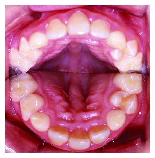
Figure 7.

Intraoral photograph with a mirror placed between the dental arches, demonstrating the palate. The figure demonstrates a single central incisor, absence of papilla incisive, and a vault midaxially in the palate. Deviations all occur within the frontonasal field, illustrated schematically in Figure 5. The figure is reprinted with permission from Neuropediatrics 2009;40:280-283 [8].
