
Figure 3. Effect of SKCa/IKCa activation on spatial decay of electrical conduction.
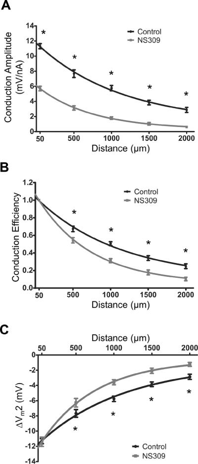
Summary data (means ± S.E.) illustrating electrical conduction versus distance between intracellular microelectrodes before and during treatment with NS309 (1 μmol/L). At each distance, continuous (paired) recordings were obtained under Control conditions and during NS309. A, For Conduction Amplitude (−1 nA microinjected at Site 1), NS309 reduced the local response by half and λ by ~40% (Control: 1380±80 μm; NS309: 850±60 μm). B, Conduction Efficiency = data from A normalized to respective values at 50 μm before and during NS309; note greater decay with NS309. C, With current microinjection adjusted to produce the same local ΔVm (Control: −1 nA; NS309: −2 nA), the ΔVm2 (= resting Vm - peak response Vm) with distance indicates greater decay of hyperpolarization with NS309. For these experiments, n = 11 at 50 – 1,500 μm; n=7 at 2,000 μm). *Control significantly different from NS309, P < 0.05
Note: Panel A includes (with permission: British Journal of Pharmacology © 2011) control data from 8 experiments presented in Figure 2B,C of Behringer et al.30