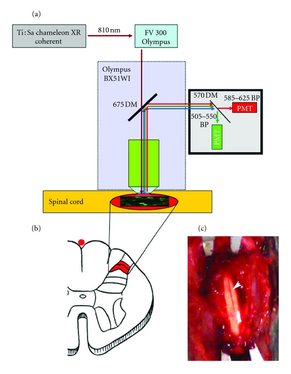
Figure 1.

Experimental setup. (a) Two-photon laser scanning microscope setup. (b) Schematic section of the lumbar spinal cord showing the regions of interest (laminae I-II). (c) Exposed spinal cord surface, after vertebral laminectomy, before SR101/OGB loading. In the middle line, the posterior medial spinal vein.
