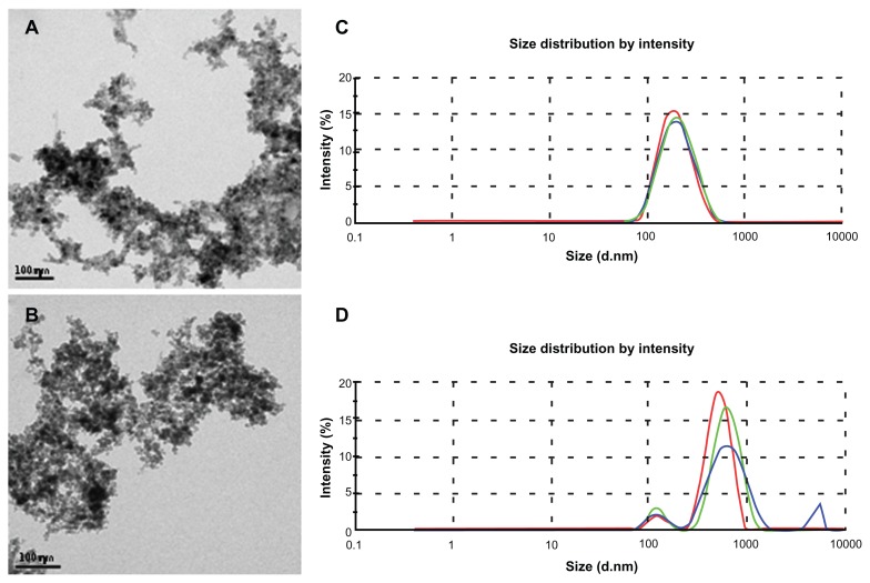
Figure 2.
Characterization of nanoparticles by transmission electron microscopy and dynamic light scattering. Electron micrograph of maghemite nanoparticles in MF sample (A) and anti-CEA conjugated nanoparticles in MF-anti-CEA sample (B). Hydrodynamic size of nanoparticles in MF sample (C) and MF-anti-CEA sample (D).
Note: Red, green, and blue lines represent the first, the second, and the third measurement of the triplicate, respectively.
Abbreviations: MF, magnetic fluid; CEA, carcinoembryonic antigen; (d · nm), diameter in nanometers.

