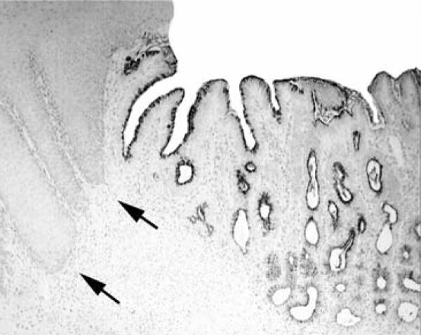
Figure 3.

Columnar-lined glandular mucosa of the esophagus in a baboon, showing sialomucin-positive cells, both along the surface epithelium as well as in the epithelium of the glands. In contrast, the squamous epithelium (arrows) remained unstained (Alcian blue, pH 2.5, without counterstain, ×4).
