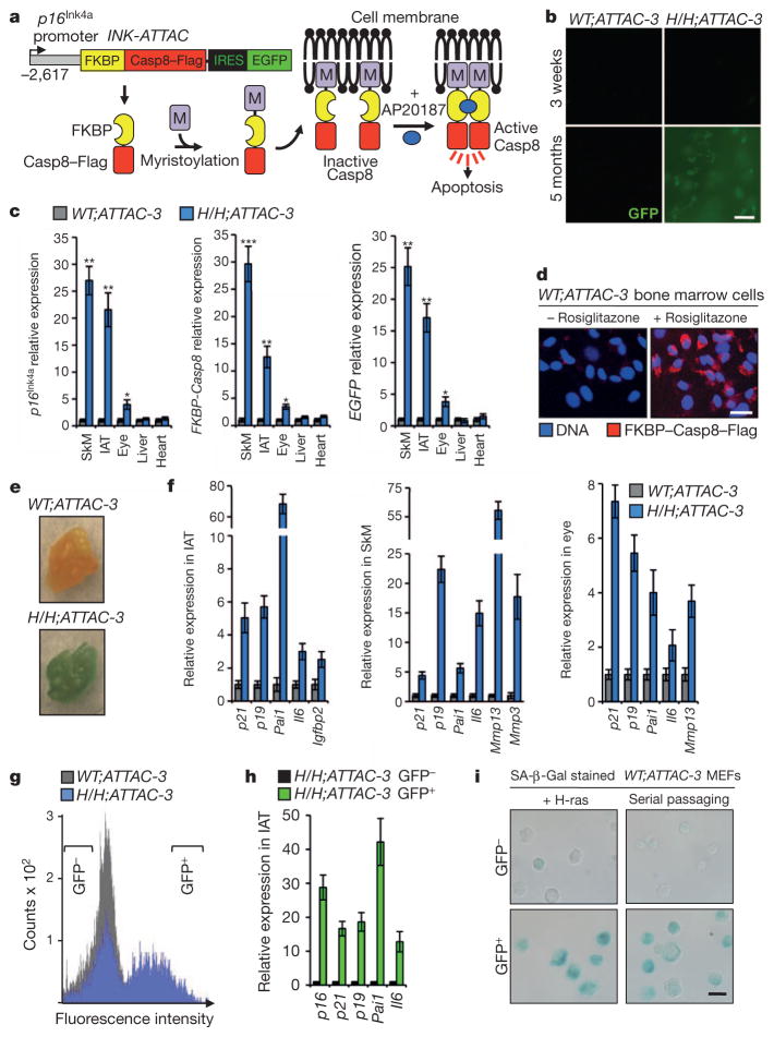
Figure 1. Generation and characterization of INK-ATTAC transgenic mice.
a, Schematic of the INK-ATTAC construct and the mechanism of apoptosis activation. b, GFP intensity of IAT. c, qRT–PCR analysis of the indicated tissues of 10-month-old mice. ATTAC, INK-ATTAC; H/H, BubR1H/H; SkM, skeletal muscle (gastrocnemius). d, Bone marrow cells harvested from 2-month-old mice immunostained for Flag after culture in the absence or presence of rosiglitazone for 48 h. e, SA-β-Gal stained IAT collected from 9-month-old mice of the indicated genotypes. f, Expression of senescence markers in tissues of 10-month-old mice measured by qRT–PCR. All increases are statistically significant (P < 0.05). g, FACS profile of single-cell suspensions from IAT of 10-month-old mice. Brackets indicate sorting gates. h, GFP+ and GFP− cell populations from IAT analysed for relative expression of senescence markers by qRT–PCR. All increases are statistically significant (P < 0.01). i, Bright field images of MEFs sorted into GFP+ and GFP− populations after induction of senescence and then stained for SA-β-Gal. For all experiments, n = 3 untreated females per genotype. Error bars, s.d. Scale bars in b, d and i, 20 μm. *P < 0.05, **P < 0.01, ***P < 0.001.

