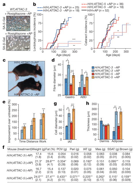
Figure 2. BubR1H/H;INK-ATTAC mice treated with AP20187 from weaning age on show delayed onset of p16Ink4a-mediated age-related phenotypes.
a, Bone marrow cells cultured in rosiglitazone for 5 days and then treated or not treated with AP20187 (AP) for 2 days before SA-β-Gal staining. Scale bar, 50 μm. b, Incidence of lordokyphosis and cataracts. c, Representative images of 9-month-old mice. d, Mean skeletal muscle fibre diameters of 10-month-old mice. ABD, abdominal muscle; Gastro, gastrocnemius muscle. e, Exercise ability of 10-month-old AP20187-treated mice relative to age-matched untreated mice. Time is running time to exhaustion; distance is distance travelled at time of exhaustion; work is the energy expended to exhaustion. f, Body and fat depot weights of 10-month-old mice. Parentheses, s.d. Mes, mesenteric; Peri, perirenal; POV, paraovarian; SSAT, subscapular adipose tissue. g, Average fat cell diameters in IAT of 10-month-old mice. h, Dermis and subdermal adipose layer thickness of 10-month-old mice. Colour codes in e, g and h are as indicated in d. Error bars, s.e.m. For all analysis n = 6 female mice per genotype (per treatment). *P < 0.05, **P < 0.01, ***P < 0.001.

