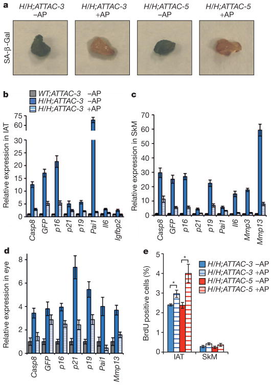
Figure 3. AP20187-treated BubR1H/H;INK-ATTAC mice have reduced numbers of p16Ink4a-positive senescent cells.
a, Images of SA-β-Gal stained IAT of 10-month-old mice. b–d, Expression of senescence markers in IAT (b), gastrocnemius (c) and eye (d) of 10-month-old AP20187-treated and untreated BubR1H/H;INK-ATTAC-3 mice relative to age-matched untreated WT;INK-ATTAC-3 mice. Error bars indicate s.d.; n = 3 females per genotype per treatment. The expression of all genes is significantly decreased upon AP20187 treatment (P < 0.05) with the exception of GFP in the eye. e, BrdU incorporation rates in IAT and skeletal muscle. Error bars, s.e.m.; n = 6 females per genotype per treatment. *P < 0.05.

