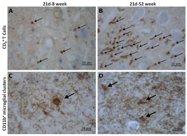
Figure 2.
Immunohistochemistry for CD3+ T lymphocyte and CD11b+ perineuronal microglial phagocytic clusters at day 21 post-resection of the facial nerve in 8 week versus 52 week old mice. CD3+ T lymphocytes and CD11b+ perineuronal microglial phagocytic clusters were immunostained (brown) and are indicated by arrows in facial motor nuclei of 8 week (A,C) and 52 week (B,D) old mice. Neuronal and glial cell bodies were counterstained with cresyl violet (blue). Incubation with either the primary antibodies or secondary antibody alone produced no signal. Scale bar = 20 μm.

