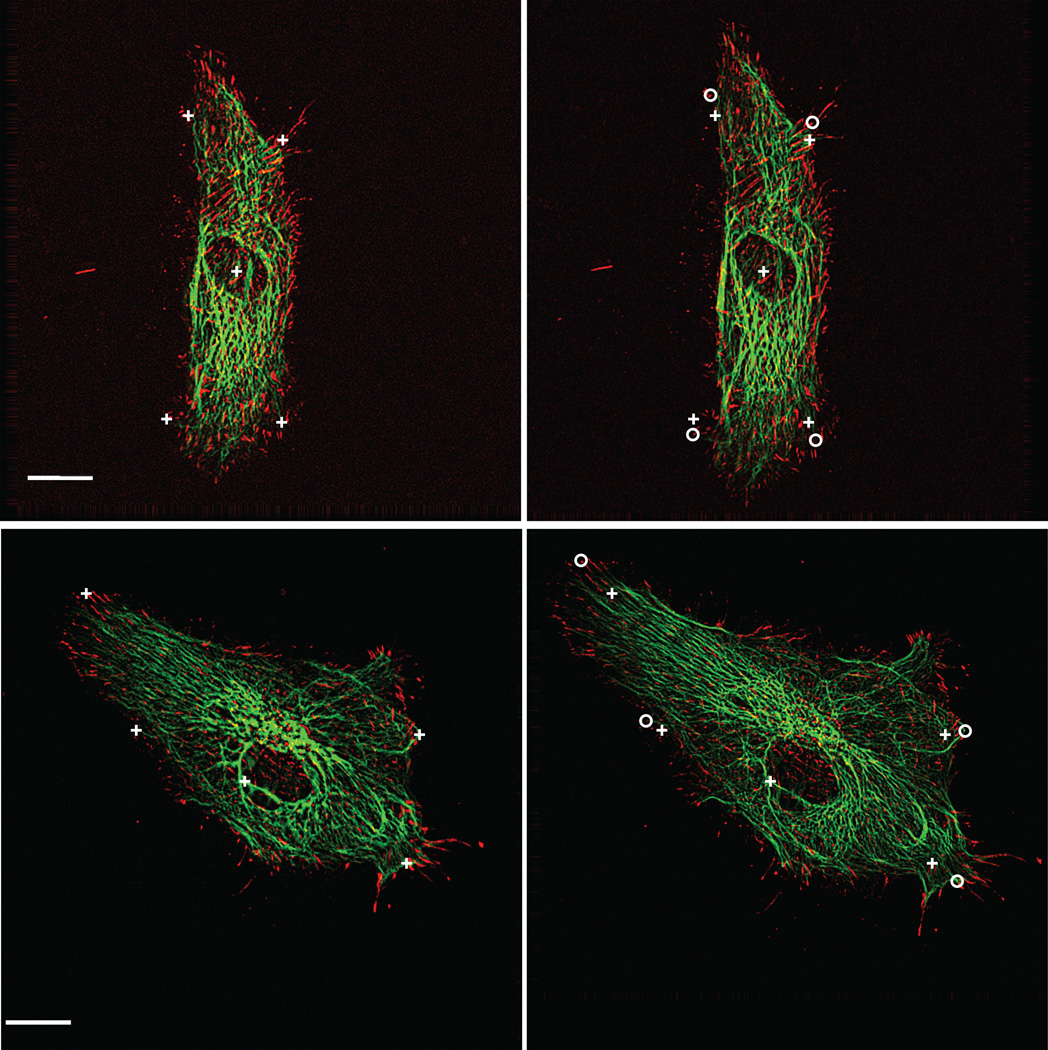
Figure 5.
Bovine aortic endothelial cells transiently expressing EGFP-vimentin (green) and paxillin-DsRed2 (red). Image pairs were acquired before and 3 min after application of 14% constant magnitude equibiaxial stretch. Adjacent cells in confluent monolayer are not expressing fluorescently labeled proteins. Markers at cell edges before (crosses) and after (circles) stretch show the extent of deformation. Center cross denotes reference fiducial used for image registration. Scale bar, 10 µm.

